
DIGITAL PATHOLOGY
Virtual staining turns a single H&E slide into quantitative biomarker data.
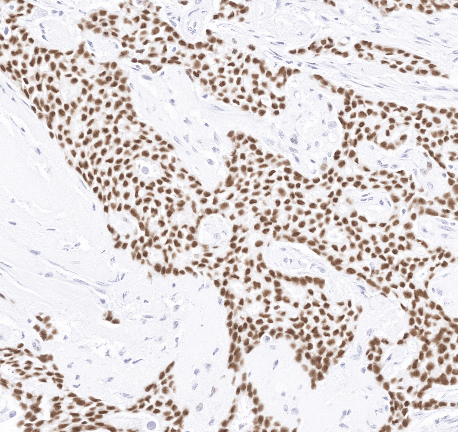
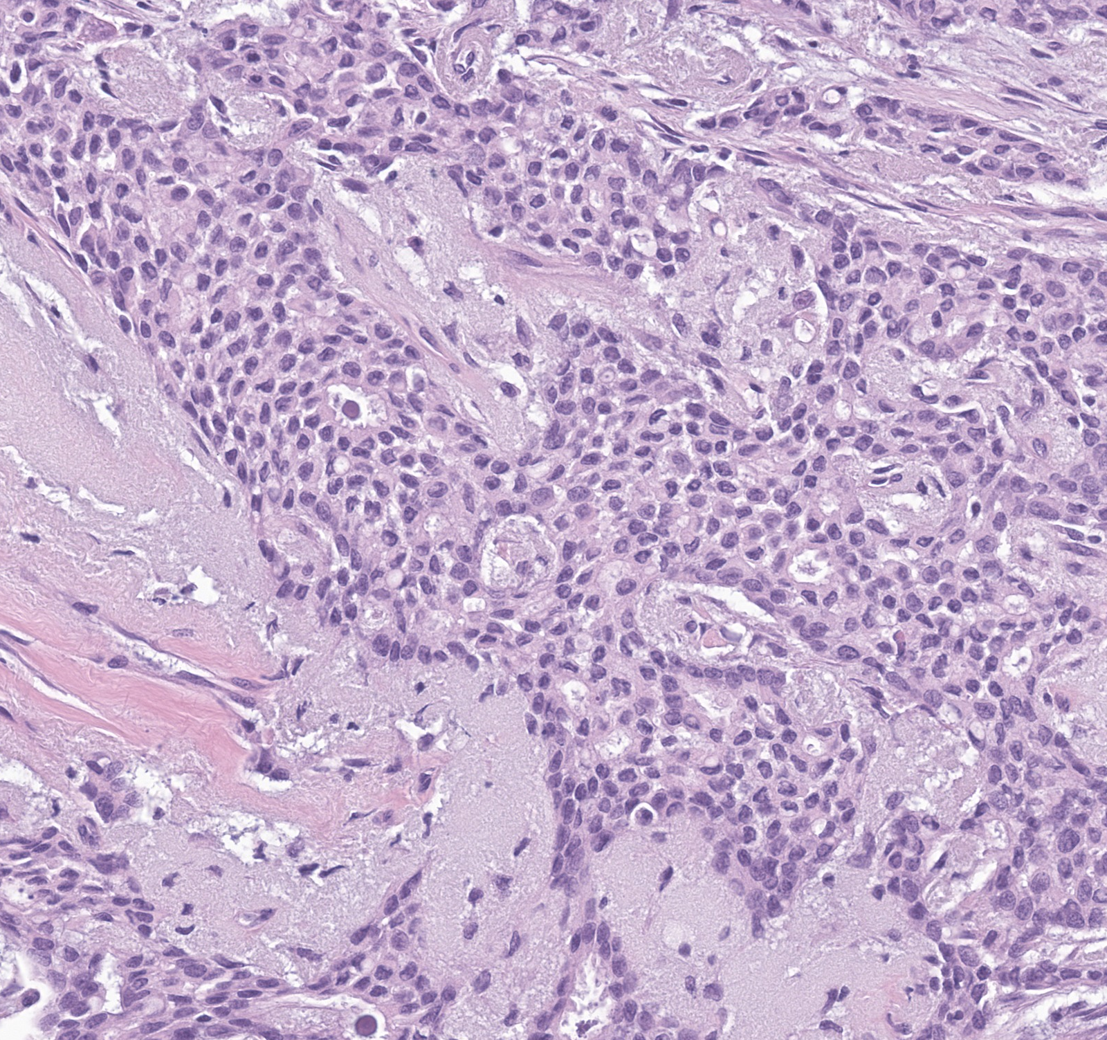
Estrogen Receptor (ER), breast carcinoma
Drag, or use the arrow keys, to compare the H&E whole slide image with the virtual stain for ER.
Obtain spatially resolved protein biomarker expression at the cell level, directly from an H&E whole slide image. No additional tissue or reagents required.
THE SHIFT
Weeks of wet-lab-intensive and tissue-consuming IHC replaced with virtual staining.
Tissue-based biomarker identification using immunohistochemistry (IHC) can take weeks, and every additional biomarker draws on already limited tissue.
Virtual staining predicts spatially resolved biomarker expression and generates the IHC image computationally from a routine H&E slide. Results are delivered in minutes, without additional tissue.
Input
One H&E slide
Turnaround
Weeks → minutes
Output
Single or multiplex IHC
ADVANTAGE
Use virtual staining to answer more biomarker questions than your tissue can supply.
Start from H&E
Use the whole slide images you already generated as part of your workflow.
Preserve tissue
Conserve tissue for downstream molecular assays, confirmatory testing, and future studies.
Multiplex from one section
Skip wet-lab multiplex IHC protocol development. Obtain results within minutes.
Unlock per-cell insight
Access per-cell spatial localization and quantitative expression data across the full slide.
Who is it for
Built for teams that need more from every tissue sample.
Translational Research
→Get spatial and quantitative biomarker data across studies, without exhausting tissue.
Clinical Trials
→Screen and stratify patients with per-cell biomarker data read directly from the routine H&E slide.
Pathology Workflow
→Building toward faster patient identification, leaner lab workflows, and less pressure on tissue, reagents, and staff.
Tissue Banking
→Extract more biomarker data from every archived block, without depleting the specimen.
GET STARTED
See virtual staining on your own tissue.
Send us one H&E whole slide image. We return the virtual IHC alongside it.
Tell us
The marker and tissue you are working on.
Send
One H&E whole slide image.
Review
Walk through the virtual IHC with our team.